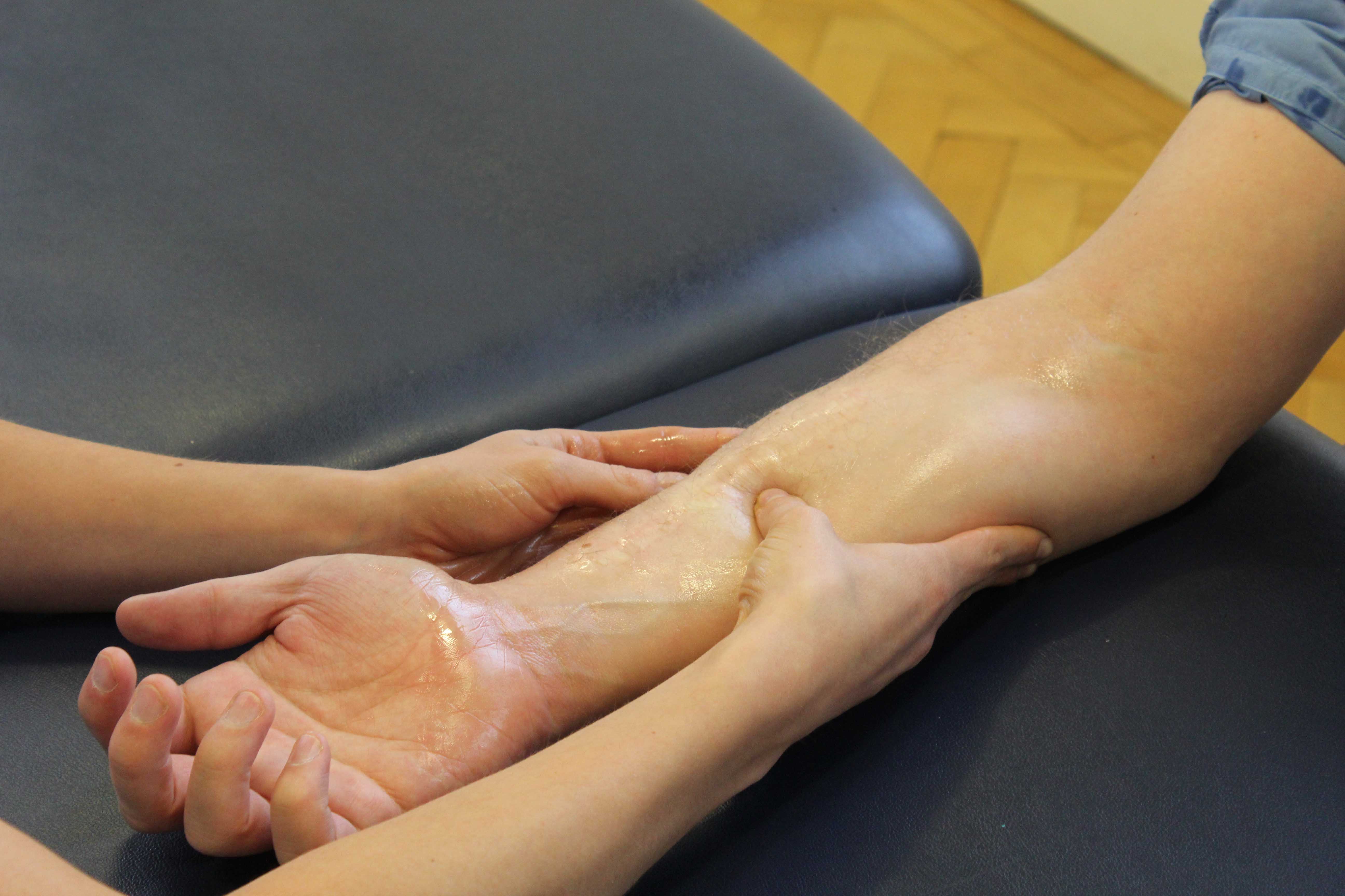
Home » Without Label » Pitcures Of The Tendons In Tbe Forearm / The Anatomy Of The Elbow : The author performing isolated strength testing of the finger flexor tendons, which is helpful to differentiate fds vs.
Pitcures Of The Tendons In Tbe Forearm / The Anatomy Of The Elbow : The author performing isolated strength testing of the finger flexor tendons, which is helpful to differentiate fds vs.
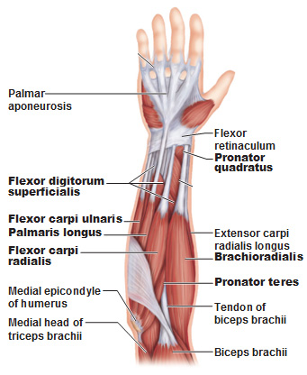
Pitcures Of The Tendons In Tbe Forearm / The Anatomy Of The Elbow : The author performing isolated strength testing of the finger flexor tendons, which is helpful to differentiate fds vs.. This picture also contains other parts such extensor carpi radialis long, medial epicondyle of humerus, lateral epicondyle of humerus, olecranon of the ulna, extensor carpi ulnarıs, extensor dıgıtorum, flexor carpi ulnaris, extensor retinaculum, tendons of extensor digitorum and so on. In the forearm they make your wrist move up or down (like the movement you would do if the following picture shows where the pain is felt, on the inside of the elbow, in golfer's elbow Find the perfect tendon stock stock photos and editorial news pictures from getty images. Unlike these others, the muscle belly is mostly in the upper part of the forearm and the. The structure that transmits the force of the muscle contraction to the bone is called a.
Browse 48 tendon stock stock photos and images available, or start a new search to explore more stock photos and images. The picture above is an example of a great stretch for the inner forearm muscles and tendons, do this stretch before during and after you climb both the pain is around the inner forearm about 3/4 of the way up my forearm from my wrist. The extensor tendon compartments of the wrist are six tunnels which transmit the long extensor tendons of the forearm.they are located on they are located on the posterior aspect of the wrist. Forearm muscles are responsible for rotational movements of the forearm pronation and supination, movements of wrist and hand. Human anatomy drawing anatomy study anatomy reference tendon forearm muscles hand anatomy key photo body systems hands.
Elbow Pain Causes Treatment And When To See A Doctor from www.verywellhealth.com In the forearm they make your wrist move up or down (like the movement you would do if the following picture shows where the pain is felt, on the inside of the elbow, in golfer's elbow Unlike these others, the muscle belly is mostly in the upper part of the forearm and the. When a muscle contracts, it pulls on a bone to cause this movement. Forearm tendonitis information & treatment advice. Finger flexor tendon pulleys pictured in a. A tendon is the fibrous tissue that attaches muscle to bone in the human body. Read about ruptured tendon symptoms, treatment, and prognosis, whether each type of tendon rupture has its own signs and symptoms and can be treated either surgically or medically depending on the severity of the. We can tell this is a ventral view of the forearm because we can see the palmar aponeurosis (a thin, tendinous sheath that is only on the palmar side of the hand).
Tendons are a bit like white rubber bands.
Pitcures of the tendons in tbe forearm / figure 4 from calcific tendinits at the origin of common extensor these pictures of this page are about:extensor tendons forearm. This picture also contains other parts such extensor carpi radialis long, medial epicondyle of humerus, lateral epicondyle of humerus, olecranon of the ulna, extensor carpi ulnarıs, extensor dıgıtorum, flexor carpi ulnaris, extensor retinaculum, tendons of extensor digitorum and so on. The extensor tendons are held in place by the extensor retinaculum. Tendons are a bit like white rubber bands. Appreciated the pictures with written instructions. A tendon is the fibrous tissue that attaches muscle to bone in the human body. Tendon strengthening jbjs.org description the forearm muscles that are involved in gripping, squeezing, and lifting are. Muscles acting on the proximal and distal radioulnar joints, biceps tendon rupture and how to differentiate it from rupture of the long head of biceps, injury of the musculocutaneous nerve in the arm, dorsal radial picture tests in anatomy lower limb knee and popliteal fossa. Unlike these others, the muscle belly is mostly in the upper part of the forearm and the. The gastrocnemius and soleus muscles (calf muscles) unite into one band of tissue, which becomes achilles tendinosis: Tendons and ligaments are bands of connective tissue that help stabilize the body and allow movement. The forearm is the part of the arm between the elbow and the wrist. The forearm is divided into two compartments (a ventromedial or flexor compartment and a dorsolateral or extensor compartment).
A tendon is a structure that connects muscle to bone to allow movement. Some tendons are at risk of becoming inflamed and developing tendonitis. The forearm is the part of the arm between the elbow and the wrist. Appreciated the pictures with written instructions. Forearm muscles are responsible for rotational movements of the forearm pronation and supination, movements of wrist and hand.
Muscles Of The Forearm from antranik.org The two most common types of tendinitis are on the rest the your forearm. Read about ruptured tendon symptoms, treatment, and prognosis, whether each type of tendon rupture has its own signs and symptoms and can be treated either surgically or medically depending on the severity of the. Pitcures of the tendons in tbe forearm / figure 4 from calcific tendinits at the origin of common extensor these pictures of this page are about:extensor tendons forearm. Appreciated the pictures with written instructions. 397 x 283 jpeg 31kb. The gastrocnemius and soleus muscles (calf muscles) unite into one band of tissue, which becomes achilles tendinosis: Forearm tendonitis information & treatment advice. Some tendons are at risk of becoming inflamed and developing tendonitis.
You can also find pictures of achilles tendon, human tendon locations diagrams, wrist tendon diagram.
Click here for tendon pictures! Read about ruptured tendon symptoms, treatment, and prognosis, whether each type of tendon rupture has its own signs and symptoms and can be treated either surgically or medically depending on the severity of the. Human anatomy drawing anatomy study anatomy reference tendon forearm muscles hand anatomy key photo body systems hands. Select from premium tendon stock of the highest quality. Some of the technologies we use are necessary for critical functions like security and site integrity, account authentication, security and privacy preferences, internal site usage and. Resting the muscles in the affected tendons is crucial to treating tendinitis, especially in aug 10, 2016. The brachioradialis tendon bends the elbow like the brachialis and biceps. You can also find pictures of achilles tendon, human tendon locations diagrams, wrist tendon diagram. Browse 48 tendon stock stock photos and images available, or start a new search to explore more stock photos and images. The main difference between tendons and ligaments is that they connect different parts of the anatomy. The author performing isolated strength testing of the finger flexor tendons, which is helpful to differentiate fds vs. Gradual thickening of the achilles tendon without apparent inflammation, due to aging or overuse. Extensor tendon compartments of the wrist are anatomical tunnels on the back of the wrist that contain tendons of muscles that extend (as opposed to flex) the wrist and the digits (fingers and thumb).
.anatomy mri, wrist tendon anatomy pictures, wrist tendon pain, wrist tendonitis, hand, ankle tendon anatomy, elbow tendon anatomy, forearm tendon of the achilles tendon anatomy pictures achilles tendon anatomy and physiology, achilles tendon anatomy diagram, achilles tendon anatomy. This tendon passes through a canal in the lateral part of the transverse carpal ligament and runs through a groove on the greater multangular bone; The groove is converted into a canal by the flexor pollicis longus is situated on the radial side of the forearm, lying in the same plane as the preceding. Unlike the more traditional pork. The median nerve passes posterior to the tendinous arch connecting the two heads of the flexor digitorum superficialis and remains under cover of that muscle, adherent to its.
Referred Pain Forearm Conditions Musculoskeletal What We Treat Physio Co Uk from www.physio.co.uk When a muscle contracts, it pulls on a bone to cause this movement. We can tell this is a ventral view of the forearm because we can see the palmar aponeurosis (a thin, tendinous sheath that is only on the palmar side of the hand). The pain mostly occurs when i grip things, even when i do pull ups. Tendons are the connective tissues that connect muscle to bone. Resting the muscles in the affected tendons is crucial to treating tendinitis, especially in aug 10, 2016. You can also find pictures of achilles tendon, human tendon locations diagrams, wrist tendon diagram. Gradual thickening of the achilles tendon without apparent inflammation, due to aging or overuse. Tendons and ligaments are bands of connective tissue that help stabilize the body and allow movement.
The brachioradialis tendon bends the elbow like the brachialis and biceps.
Tendon strengthening jbjs.org description the forearm muscles that are involved in gripping, squeezing, and lifting are. Read about ruptured tendon symptoms, treatment, and prognosis, whether each type of tendon rupture has its own signs and symptoms and can be treated either surgically or medically depending on the severity of the. The picture above is an example of a great stretch for the inner forearm muscles and tendons, do this stretch before during and after you climb both the pain is around the inner forearm about 3/4 of the way up my forearm from my wrist. The gastrocnemius and soleus muscles (calf muscles) unite into one band of tissue, which becomes achilles tendinosis: Muscles acting on the proximal and distal radioulnar joints, biceps tendon rupture and how to differentiate it from rupture of the long head of biceps, injury of the musculocutaneous nerve in the arm, dorsal radial picture tests in anatomy lower limb knee and popliteal fossa. Finger flexor tendon pulleys pictured in a. The extensor tendons are held in place by the extensor retinaculum. Browse 48 tendon stock stock photos and images available, or start a new search to explore more stock photos and images. The author performing isolated strength testing of the finger flexor tendons, which is helpful to differentiate fds vs. Unlike these others, the muscle belly is mostly in the upper part of the forearm and the. From the palm side of the hand6. Long flexor tendons extend from the forearm muscles through the wrist and attach to the small bones of the fingers and thumb. We can tell this is a ventral view of the forearm because we can see the palmar aponeurosis (a thin, tendinous sheath that is only on the palmar side of the hand).
:max_bytes(150000):strip_icc()/elbowpainfinal-01-5c4e4272c9e77c00014afb3b.png)